
CO:05:3 | Radiomics and machine learning for differentiating rheumatoid arthritis-associated interstitial lung disease and idiopathic pulmonary fibrosis with usual interstitial pneumonia pattern
Vincenzo Venerito1, Chiara Nani2, Andreina Manfredi3, Marco Fornaro1, Giuseppe Lopalco1, Florenzo Iannone1, Cecilia Burattini2, Marco Sebastiani4. | 1Rheumatology Unit, University of Bari; 2Respiratory Unit, Guglielmo da Saliceto Civil Hospital, Piacenza; 3Rheumatology Unit, Azienda Unità Sanitaria Locale-IRCCS di Reggio Emilia; 4Rheumatology Unit, AUSL Piacenza, University of Parma, Italy
All claims expressed in this article are solely those of the authors and do not necessarily represent those of their affiliated organizations, or those of the publisher, the editors and the reviewers. Any product that may be evaluated in this article or claim that may be made by its manufacturer is not guaranteed or endorsed by the publisher.
Authors
Background. While Idiopathic Pulmonary Fibrosis (IPF) and Rheumatoid Arthritis-associated Interstitial Lung Disease (RA-ILD) can manifest identical Usual Interstitial Pneumonia (UIP) patterns on HRCT, their underlying histopathology reveals fundamentally different disease processes—IPF driven by aberrant fibroblast proliferation and excessive collagen deposition versus RA-ILD's autoimmune-mediated inflammation. This pathological divergence translates to dramatically different prognoses and treatment responses, yet current imaging interpretation cannot reliably capture these microscopic differences. Radiomics offers a revolutionary approach: extracting hundreds of quantitative features that may encode histopathological signatures within standard HRCT images. Combined with machine learning, this technology might unveil tissue-level distinctions invisible to visual analysis, potentially transforming a diagnostic challenge into a data-driven classification. Our aim was to develop a radiomic-based framework for distinguishing IPF from RA-ILD in UIP-pattern patients.
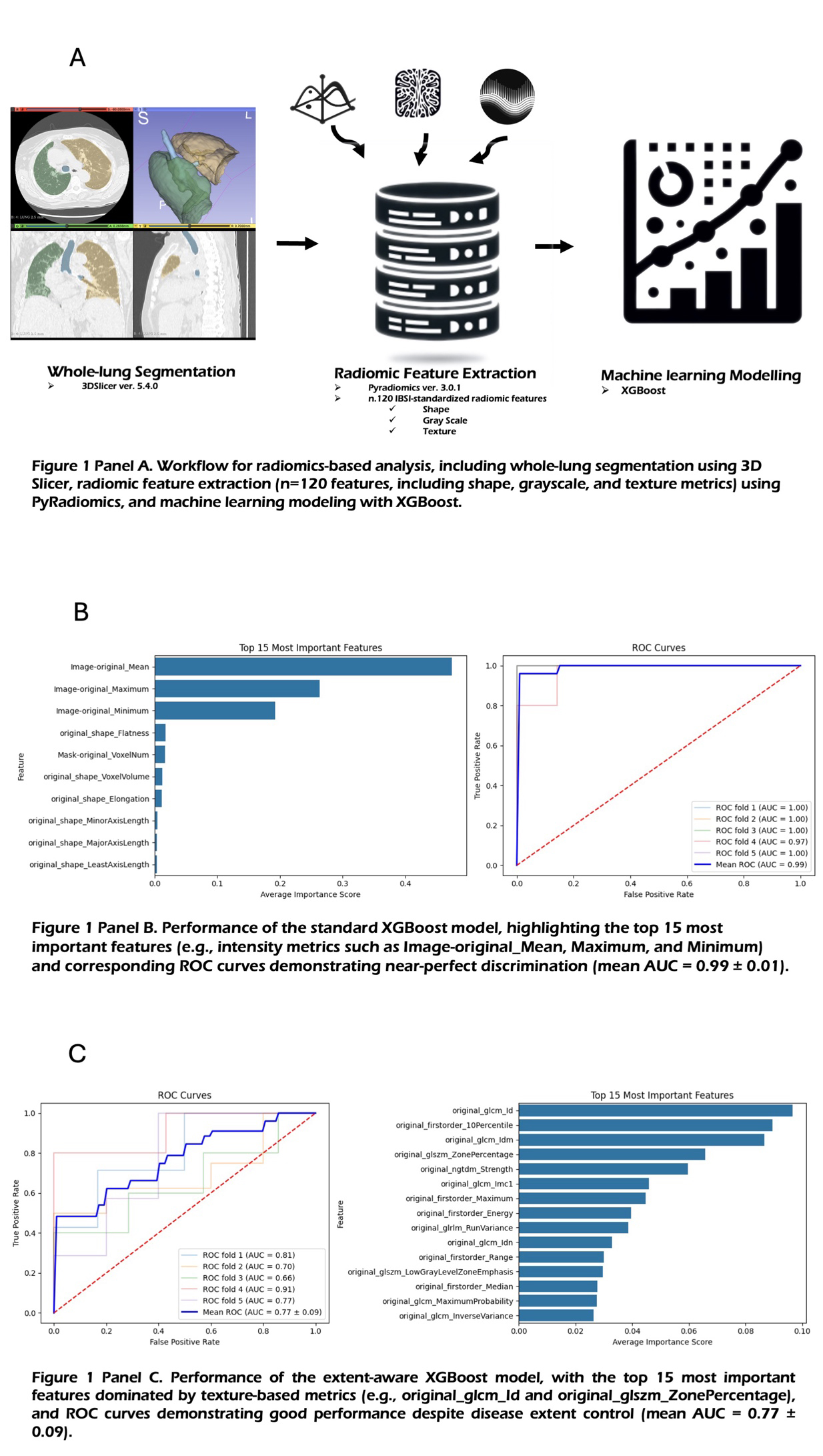
Methods. We conducted a retrospective analysis of patients with confirmed UIP pattern diagnosed in tertiary centres. Whole-lung volumes were semi-automatically segmented using 3DSlicer (v5.4.0, https://tinyurl.com/mr339kmm). 120 radiomic features were extracted via PyRadiomics (v3.0.1). Features underwent z-score standardization and multicollinearity assessment Two distinct XGBoost models were developed to address different clinical scenarios: Standard Model: Leveraged all radiomic features to maximize discriminative performance. - Extent-Aware Model: Excluded volume-related features and incorporated mesh volume as a covariate to control for disease severity, ensuring classification based on intrinsic tissue characteristics rather than disease burden. Model performance was evaluated using 5-fold cross-validation with area under the receiver operating characteristic curve (AUC) as the primary metric.
Results. The study included 72 patients: 42 with IPF (male 73.8%, median age 78, IQR 8) and 30 with RA-ILD (female, 63.3%, median age 72. IQR 12.5). Standard Model achieved near-perfect discrimination (AUC 0.99±0.01), driven by first-order intensity features: Image-original_Mean (overall lung density); Image-original_Maximum/Minimum (attenuation extremes); Extent-Aware Model maintained robust performance (AUC 0.77±0.09) using texture-based features: original_glcm_Id (texture homogeneity); original_firstorder_10Percentile (lower intensity distribution); original_glszm_ZonePercentage (homogeneous zone proportion) IPF median survival 5.5 months versus 29.5 months for RA-ILD.
Conclusions. Radiomics with machine learning demonstrates exceptional potential for differentiating IPF from RA-ILD in UIP-pattern patients. The near-perfect accuracy suggests quantitative biomarkers capture disease-specific signatures beyond visual assessment. Crucially, the extent-aware model's success indicates fundamental tissue architecture differences between conditions, independent of disease severity. Implementing radiomic analysis could enable earlier diagnosis and timely disease-specific therapy. 
Downloads
Citations


How to Cite

This work is licensed under a Creative Commons Attribution-NonCommercial 4.0 International License.


