
62nd National Congress of the Italian Society of Rheumatology
Vol. 77 No. s1 (2025): Abstract book of the 62th Conference of the Italian Society for...
PO:33:202 | Assessment of Retinal Arteriolar Wall-to-Lumen Ratio and Renal Resistive Index in patients with Systemic Sclerosis
Eleonora Pazzi1|2, Elda Piovani1|2, Claudia Barison1|2, Liala Moschetti1|2, Eleonora Pedretti1|2, Mariagrazia Lazzaroni1|2, Carolina De Ciuceis3, Claudia Agabiti-Rosei3, Giacomo Buso3|4, Paolo Baggi3, Claudia Rossini3, Cristina Bevacqua3, Maria Lorenza Muiesan3, Ilaria Cavazzana1, Franco Franceschini1, Paolo Airò2 | 1Rheumatology and Clinical Immunology, ASST Spedali Civili of Brescia, University of Brescia, Brescia, Italy; 2Scleroderma Unit, ASST Spedali Civili of Brescia, Brescia, Italy; 3Department of Clinical and Experimental Sciences, Division of Internal Medicine, ASST Spedali Civili of Brescia, Univers Brescia, Italy; 4University of Lausanne, Lausanne Switzerland
Publisher's note
All claims expressed in this article are solely those of the authors and do not necessarily represent those of their affiliated organizations, or those of the publisher, the editors and the reviewers. Any product that may be evaluated in this article or claim that may be made by its manufacturer is not guaranteed or endorsed by the publisher.
All claims expressed in this article are solely those of the authors and do not necessarily represent those of their affiliated organizations, or those of the publisher, the editors and the reviewers. Any product that may be evaluated in this article or claim that may be made by its manufacturer is not guaranteed or endorsed by the publisher.
Published: 18 March 2026
45
Views
Authors
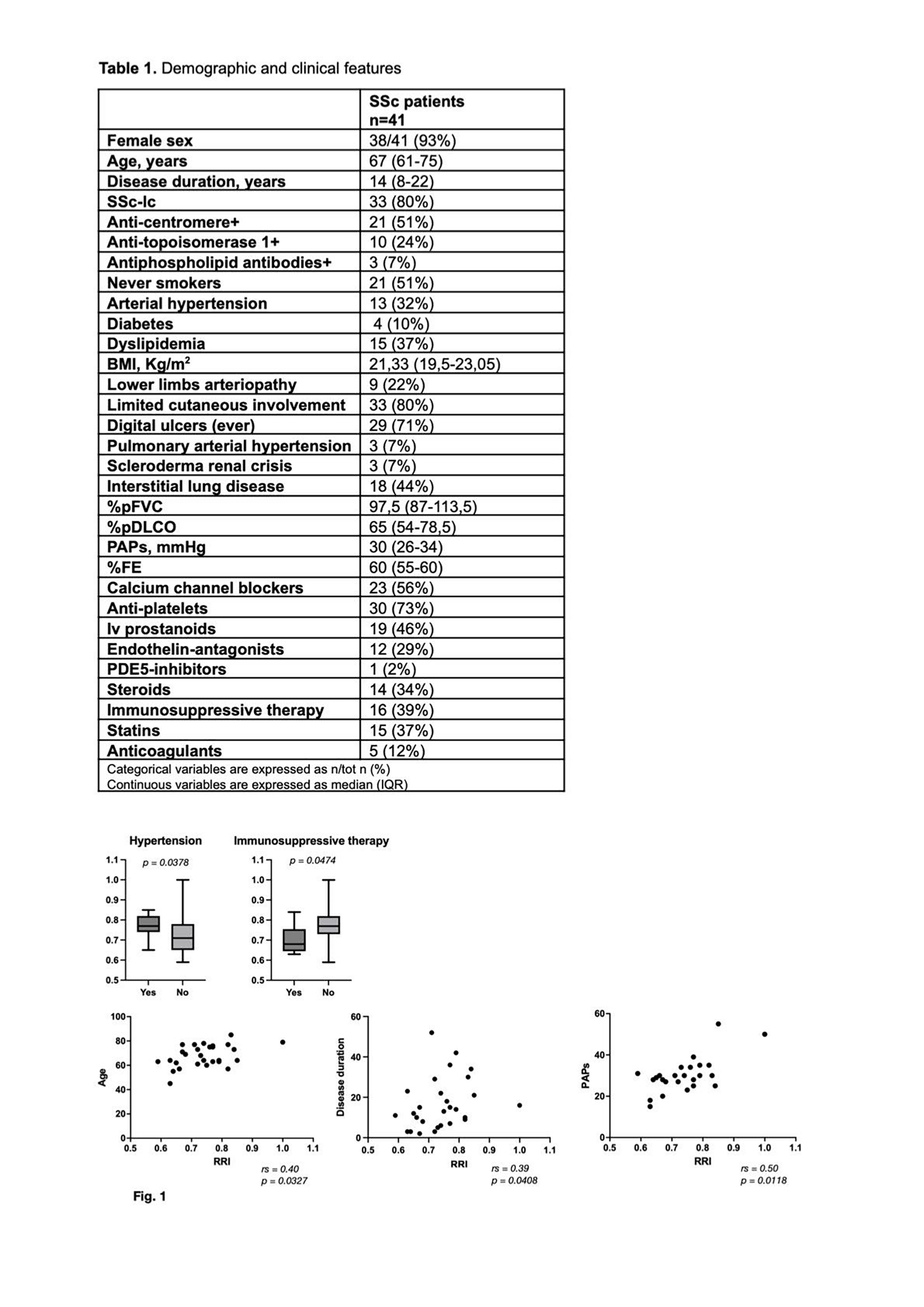
Materials and Methods. Forty-one SSc patients were enrolled in this study. The following examinations were proposed to all patients: Non-invasive evaluation of retinal arterioles by Adaptive Optics. Kidney arteries DUS. Lower extremity arterial DUS. Statistical comparisons were performed using the Mann–Whitney U test, while correlations were evaluated using Spearman’s rank correlation coefficient (rs)
Results. Demographic and clinical features of included SSc patients are reported in Table 1. Completed assessments included: retinal WLR determination (n=24), kidney (n=28) and lower extremity DUS (n=25). Median WLR was 0.25 (0.23-0.27), with only 5 patients (21%) showing values greater than 0.28. Median renal resistivity index (RRI; normal values <0.70) was 0.74 (0.67-0.79) in the right kidney; 19 (68%) patients presented RRI over 0.70. DUS of the lower limbs showed the presence of peripheral arterial disease in 9 (36%) patients. No significant differences in WLR values were observed when stratifying the cohort by the analyzed parameters. RRI was increased in patients with arterial hypertension (p=0.0378) and in patients not treated with immunosuppressive therapy (p=0.047). Moreover, RRI showed a positive correlation with age (rs=0.40; p=0.032), disease duration (rs=0.39; p=0.041), and estimated PAPs on cardiac ultrasound (rs=0.50; p=0.012) (Fig. 1). No significant correlation emerged between WLR and RRI (rs=-0.10; p=0.689).
Conclusions. Retinal WLR was increased only in a relatively small number of SSc patients included in the study and does not appear to be associated with increased frequency of cardiovascular manifestations in this small cohort. The use of vasoactive drugs (calcium-channel blockers, ERA, IV prostanoids) and antiplatelet therapy in the large majority of the included patients might have affected these observations. On the other hand, kidney DUS demonstrates frequent arterial abnormalities. Further longitudinal studies on a larger number of patients with SSc may be warranted to evaluate the role of retinal arterioles and kidney arteries study in them. References 1. De Ciuceis, C. et al. European Journal of Internal Medicine, 122, 86 - 92. 2. Nathalie Conrad et al. Lancet 2022; 400: 733–43. Acknowledgements. The Authors thank GILS (Gruppo Italiano Lotta Sclerodermia) for substantially supporting this project.
Downloads
Download data is not yet available.
Citations


How to Cite
1.
PO:33:202 | Assessment of Retinal Arteriolar Wall-to-Lumen Ratio and Renal Resistive Index in patients with Systemic Sclerosis: Eleonora Pazzi1|2, Elda Piovani1|2, Claudia Barison1|2, Liala Moschetti1|2, Eleonora Pedretti1|2, Mariagrazia Lazzaroni1|2, Carolina De Ciuceis3, Claudia Agabiti-Rosei3, Giacomo Buso3|4, Paolo Baggi3, Claudia Rossini3, Cristina Bevacqua3, Maria Lorenza Muiesan3, Ilaria Cavazzana1, Franco Franceschini1, Paolo Airò2 | 1Rheumatology and Clinical Immunology, ASST Spedali Civili of Brescia, University of Brescia, Brescia, Italy; 2Scleroderma Unit, ASST Spedali Civili of Brescia, Brescia, Italy; 3Department of Clinical and Experimental Sciences, Division of Internal Medicine, ASST Spedali Civili of Brescia, Univers Brescia, Italy; 4University of Lausanne, Lausanne Switzerland. Reumatismo [Internet]. 2026 Mar. 18 [cited 2026 Jul. 27];77(s1). Available from: https://www.reumatismo.org/reuma/article/view/2377
Copyright (c) 2026 The Author(s)


This work is licensed under a Creative Commons Attribution-NonCommercial 4.0 International License.


