
62nd National Congress of the Italian Society of Rheumatology
Vol. 77 No. s1 (2025): Abstract book of the 62th Conference of the Italian Society for...
PO:32:187 | AI-assisted reading outperforms visual scoring of pulmonary CT scans to predict interstitial lung disease progression in systemic sclerosis
Francesca Motta1, Antonio Tonutti1, Federica Catapano2, Gianluca Sellaro2, Francesco Amati3, Anna Stainer3, Calogero Messana2, Stefano Erba1, Stefano Aliberti3, Marco Francone2, Carlo Selmi1, Maria De Santis1 | 1Humanitas University and IRCCS Humanitas Research Hospital, Department of Rheumatology and Clinical Immunology Rozzano, Milan, Italy; 2IRCCS Humanitas Research Hospital, Department of Radiology Rozzano, Milan, Italy; 3IRCCS Humanitas Research Hospital, Respiratory Unit Rozzano, Milan, Italy
Publisher's note
All claims expressed in this article are solely those of the authors and do not necessarily represent those of their affiliated organizations, or those of the publisher, the editors and the reviewers. Any product that may be evaluated in this article or claim that may be made by its manufacturer is not guaranteed or endorsed by the publisher.
All claims expressed in this article are solely those of the authors and do not necessarily represent those of their affiliated organizations, or those of the publisher, the editors and the reviewers. Any product that may be evaluated in this article or claim that may be made by its manufacturer is not guaranteed or endorsed by the publisher.
Published: 18 March 2026
52
Views
Authors
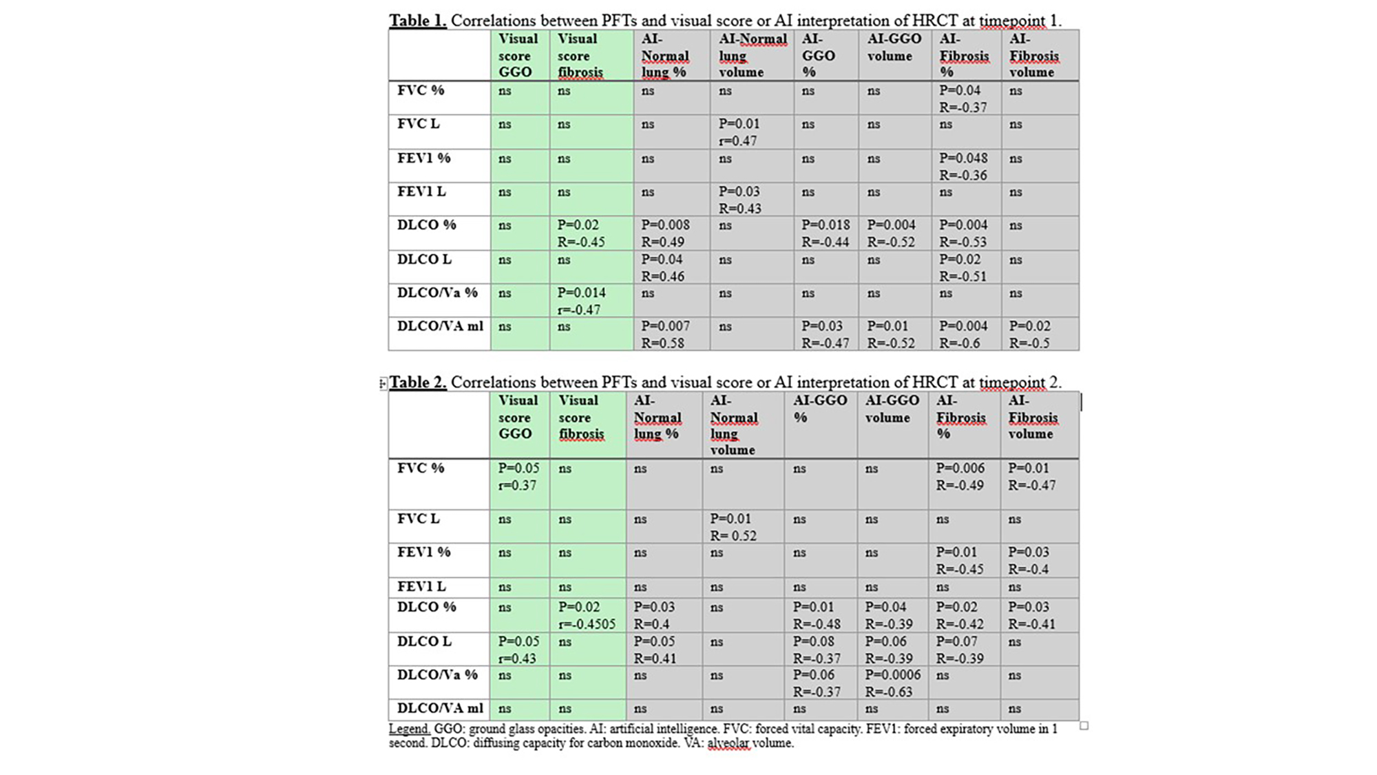
Methods. In this retrospective, longitudinal study, 33 patients with SSc-ILD (79% female, 76% anti-Scl70+, 60% on treatment, of which 75% with mycophenolate at an average dose 2.075 g/day) underwent pulmonary function tests (PFT) and HRCT scans at two timepoints 12 months apart (T1, T2). A third timepoint (T3) after an additional 12 months was available in a subset of patients (n=20). HRCT scans were assessed by two chest radiologists for semi-quantitative evaluation of fibrosis and ground-glass opacities (GGO), and by AI-assisted software (Thoracic VCAR, GE Healthcare) to evaluate and quantify volume and percentage of fibrosis, GGO, emphysema, and consolidations. ILD progression was defined using Erice functional criteria (2) and the minimal clinically important worsening (MCIW) of FVC (decline of 3% or more over 12 months).
Results. Patient showed a significant reduction of forced vital capacity (FVC)% from T1 to T2 (85 –SD 21- to 80 – SD 17- %, p=0.04), but no difference in other PFT parameters. Both visual and AI-assisted HRCT analyses showed significant increase in fibrosis scores over time (visual: 1.5 to 1.6, p=0.0005; AI: 2.2 to 2.4%, p=0.0007). AI also detected a significant increase in GGO% (15.5 to 15.8%, p = 0.04) and reduction in normal lung (75.6 to 73%, p=0.009). AI-derived HRCT parameters strongly correlated with PFT values, particularly FVC and diffusing capacity for carbon monoxide (DLCO), at both T1 and T2. In contrast, visual scores showed weaker correlations (Tables 1 and 2). Concordance between visual and AI assessments of GGO and fibrosis was low or absent at both timepoints. Progressive ILD at T3 was predicted by the increase of AI-determined volume of GGO from T1 to T2, even after adjustment for age, gender, and disease duration (P=0.04, OR 9, 95%CI 1.37-15). Visual scoring did not predict progression.
Conclusions. The AI-assisted quantitative HRCT analysis detects early SSc-ILD progression and may identify patients at risk of functional decline better and earlier than PFT.
References
1. Kazerooni EA, et al. AJR Am J Roentgenol. 1997 Oct;169(4):977-83. 2. George PM, et al. Lancet Respir Med. 2020 Sep;8(9):925-934.
Downloads
Download data is not yet available.
Citations


How to Cite
1.
PO:32:187 | AI-assisted reading outperforms visual scoring of pulmonary CT scans to predict interstitial lung disease progression in systemic sclerosis: Francesca Motta1, Antonio Tonutti1, Federica Catapano2, Gianluca Sellaro2, Francesco Amati3, Anna Stainer3, Calogero Messana2, Stefano Erba1, Stefano Aliberti3, Marco Francone2, Carlo Selmi1, Maria De Santis1 | 1Humanitas University and IRCCS Humanitas Research Hospital, Department of Rheumatology and Clinical Immunology Rozzano, Milan, Italy; 2IRCCS Humanitas Research Hospital, Department of Radiology Rozzano, Milan, Italy; 3IRCCS Humanitas Research Hospital, Respiratory Unit Rozzano, Milan, Italy. Reumatismo [Internet]. 2026 Mar. 18 [cited 2026 Jul. 27];77(s1). Available from: https://www.reumatismo.org/reuma/article/view/2373
Copyright (c) 2026 The Author(s)


This work is licensed under a Creative Commons Attribution-NonCommercial 4.0 International License.


