
62nd National Congress of the Italian Society of Rheumatology
Vol. 77 No. s1 (2025): Abstract book of the 62th Conference of the Italian Society for...
PO:07:097 | CGRP and CALCRL localization supports the role of fibroblast-like synoviocytes in rheumatoid arthritis pain pathways
Giada La Spina1, Cristina Garufi2, Mattia Caliste2, Martina Leopizzi3, Valeria Di Maio3, Cristiano Alessandri2, Fabrizio Conti2, Francesca Romana Spinelli2 | 1Sapienza University of Rome, Dipartimento di Medicina Molecolare, Roma, Italy; 2Sapienza University of Rome, Dipartimento di Scienze Cliniche Internistiche, Anestesiologiche e Cardiovascolari, Roma, Italy; 3Sapienza University of Rome, Dipartimento di Scienze e Biotecnologie Medico-Chirurgiche, Roma, Italy
Publisher's note
All claims expressed in this article are solely those of the authors and do not necessarily represent those of their affiliated organizations, or those of the publisher, the editors and the reviewers. Any product that may be evaluated in this article or claim that may be made by its manufacturer is not guaranteed or endorsed by the publisher.
All claims expressed in this article are solely those of the authors and do not necessarily represent those of their affiliated organizations, or those of the publisher, the editors and the reviewers. Any product that may be evaluated in this article or claim that may be made by its manufacturer is not guaranteed or endorsed by the publisher.
Published: 18 March 2026
45
Views
Authors
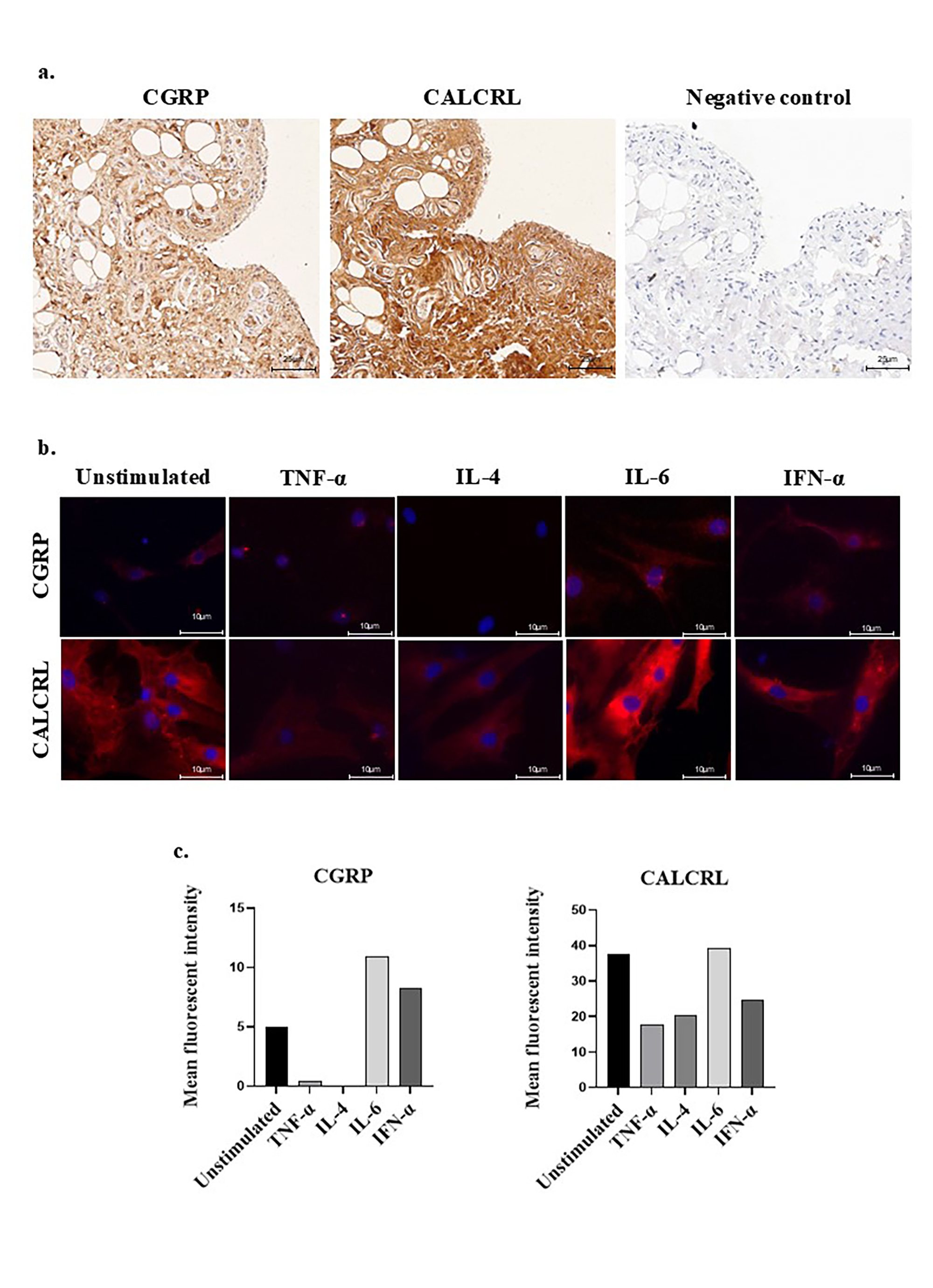
Methods. CGRP and CALCRL immunohistochemistry (IHC) was performed on synovial biopsies from 3 RA patients incubated with primary antibodies (anti-CGRP, Santa Cruz, sc-57043; anti-CALCRL, Thermo Fisher, 8H9L8; both 1:100). Positivity for CGRP and CALCRL was defined as the presence of cytoplasmic immunostaining and the intensity was graded as: negative, low, moderate, and high staining. RA-FLS were isolated from synovial fluid and cultured in complete medium. At passage 3, RA-FLS were seeded on eight-well slides, stimulated with TNF-alpha (5 ng/ml), IL-4 (5 ng/ml), IL-6 (25 ng/ml), or IFN-alpha (1000 U/ml) for 24h. Cells were fixed and then incubated with the primary antibodies (anti-CGRP, anti-CALCRL; both 1:50) and Alexa Fluor 555 tyramide (1:200). The slides were visualized with an Olympus IX50 fluorescence microscope and signal intensity was quantified using ImageJ.
Results. In synovial tissue, CGRP was moderately expressed in fibroblasts, highly abundant within blood vessel and absent in synoviocytes. Conversely, CALCRL is strongly expressed in both fibroblasts and synoviocytes, but not detected in the vessels (Figure 1a). In unstimulated RA-FLS, immunofluorescence analysis showed CALCRL and CGRP localized in the cytoplasm, with moderate and weak signal intensity, respectively. After IL-6 and IFN-alpha treatment, a moderate signal intensity for CALCRL was detected in the cytoplasm with strong perinuclear enhancement; also, a slight perinuclear increase of CGRP signal intensity was observed. In TNF-alpha and IL-4 treatment, a strong reduction was observed in the signal intensity of both CGRP and CALCRL in the cytoplasm (Figure 1b-1c).
Conclusions. These findings suggest that CGRP and its receptor CALCRL exhibit distinct expression patterns in synovial tissue. Notably, CALCRL expression is consistently higher than its ligand CGRP, indicating that both types of synovial cells may play an active role in neuro-inflammatory pain mediated by the CGRP/CALCRL pathway in RA. Furthermore, stimulation experiments on RA-FLS demonstrated that IL-6 modulates the expression of both CGRP and CALCRL , suggesting that IL-6 could be crucial in modulating this pain-related pathway in RA patients. Figure 1 a. IHC of RA synovial tissue shows CGRP and CALCRL cytoplasmic localization. Positive staining appears brown; nuclei are counterstained blue with hematoxylin. b. IF of RA-FLS after 24-hour stimulation with TNF-alpha, IL-4, IL-6 or IFN-alpha compared to unstimulated (basal) conditions. Cells are stained for CGRP and CALCRL (red); nuclei are counterstained with Hoechst (blue). c. Bar graph showing CGRP and CALCRL mean fluorescence intensity post-FLS cytokine stimulation.
Downloads
Download data is not yet available.
Citations


How to Cite
1.
PO:07:097 | CGRP and CALCRL localization supports the role of fibroblast-like synoviocytes in rheumatoid arthritis pain pathways: Giada La Spina1, Cristina Garufi2, Mattia Caliste2, Martina Leopizzi3, Valeria Di Maio3, Cristiano Alessandri2, Fabrizio Conti2, Francesca Romana Spinelli2 | 1Sapienza University of Rome, Dipartimento di Medicina Molecolare, Roma, Italy; 2Sapienza University of Rome, Dipartimento di Scienze Cliniche Internistiche, Anestesiologiche e Cardiovascolari, Roma, Italy; 3Sapienza University of Rome, Dipartimento di Scienze e Biotecnologie Medico-Chirurgiche, Roma, Italy. Reumatismo [Internet]. 2026 Mar. 18 [cited 2026 Jul. 14];77(s1). Available from: https://www.reumatismo.org/reuma/article/view/2303
Copyright (c) 2026 The Author(s)


This work is licensed under a Creative Commons Attribution-NonCommercial 4.0 International License.


