
PO:11:158 | Osteomyelitis of the jaw: a rare mimick of giant cell arteritis
Abstract
Background. Osteomyelitis of the jaw is a complication of odontogenic infections, primarily involving the dentate portion of the mandible. Osteomyelitis of the mandibular condyle is exceptionally rare, and spread may occur via hematogenous or contiguous routes (1). We here present a case of osteomyelitis of the mandibular condyle initially diagnosed as Horton's arteritis.
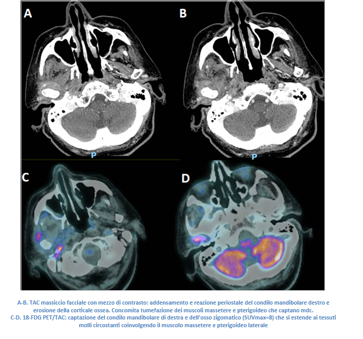
Case report. An 87-year-old woman with a recent diagnosis of giant cell arteritis underwent rheumatological evaluation (Figure 1). The patient had been complaining for 1 year of non-NSAIDs-responsive headache in the temporal and preauricular regions and jaw claudication. A few months earlier the headache onset, she underwent a tooth extraction with the positioning of a mobile prosthesis. The patient reported that she was first evaluated by a neurologist who, considering the elevation of inflammatory lab tests (CRP 2.8 mg/dL), diagnosed Horton's arteritis and started prednisone 50 mg/day, with remission of the symptoms, but relapse at steroids tapering. At the first rheumatological evaluation, the patient reported pain in the right temporal region, greater at night, with a sense of ear fullness, hyporexia, and weight loss. In laboratory tests conducted under a low-dose steroids regimen (prednisone 7.5 mg/day), a modest increase in inflammation (CRP 1.5 mg/dL) was observed. For a better diagnostic review, the steroids were discontinued, and the patient underwent a temporal artery Doppler ultrasound, which detected no arteritis-compatible abnormalities. Given the worsening of pain, the rise in inflammation (CRP 4 mg/dL) and the swelling of the soft tissues upon discontinuation of the steroids, the patient underwent an 18FDG-PET/CT scan, which excluded vascular uptake but demonstrated intense uptake of the mandibular condyle, pterygoid muscle, and zygomatic bone. She also underwent a contrast-enhanced CT scan, which revealed erosions and periosteal reaction in the jaw, suggestive of osteomyelitis, along with inflammation of the adjacent soft tissues.
Conclusions. Mandibular condyle osteomyelitis is a rare complication of dental infections and invasive dental procedures. The associated symptomatology (temporal headache, difficulty in chewing) may resemble giant cell arteritis. An accurate medical history, which also examines previous dental history and pain features, may guide the diagnosis. Anyway, radiologic exams and/or temporal artery biopsy are still necessary for differential diagnosis.

Metrics
Citations and online attention from Dimensions, Altmetric and PlumX.
License
Copyright (c) 2025 The Author(s)

This work is licensed under a Creative Commons Attribution-NonCommercial 4.0 International License.
PAGEPress has chosen to apply the Creative Commons Attribution NonCommercial 4.0 International License (CC BY-NC 4.0) to all manuscripts to be published.