Comparison of clinicodemographic characteristics and pattern of vascular involvement in 126 patients with Takayasu arteritis: a report from Iran and Turkey

Submitted: 4 March 2022
Accepted: 11 October 2022
Published: 29 December 2022
Abstract Views: 618
PDF: 307
Publisher's note
All claims expressed in this article are solely those of the authors and do not necessarily represent those of their affiliated organizations, or those of the publisher, the editors and the reviewers. Any product that may be evaluated in this article or claim that may be made by its manufacturer is not guaranteed or endorsed by the publisher.

Authors

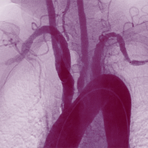
Takayasu arteritis (TA) is an extremely uncommon vasculitis that primarily affects the aorta and its branches. Due to the genetic and ethnicity effect, a diverse array of TA clinical manifestations has been reported worldwide. The purpose of the present study was to compare the clinicodemographic characteristics and pattern of vascular involvement of Iranian and Turkish TA patients. This study was a retrospective, cross-sectional investigation of 126 TA patients in Iran and Turkey. All of the variables analyzed were extracted from historical medical records. In 126 TA patients, the ratio of females to males was 8.6:1, and the average age at onset of disease was 30.5±11.1 years. Fatigue (49.2%) and a weak or absent pulse (79.4%) were the most prevalent symptoms and signs, respectively. The most prevalent angiographic classifications were types V and I in Iranian patients (41.09%) and type I in the Turkish population (47.7%) The left subclavian artery was the vessel most frequently affected by TA (66.6%). Our findings indicated that there were no significant differences between the two countries in terms of clinicodemographic characteristics or vascular involvement. Some clinical manifestations, such as claudication, were more prevalent in the Turkish population due to a higher incidence of occlusive lesions in the right subclavian artery.

Dimensions

Altmetric

PlumX Metrics

Downloads

Download data is not yet available.

Citations

Cotch M, Hoffman G, editors. The prevalence, epidemiology and cost of hospitalizations for vasculitis in New-York-State-1986 to 1990. Arthritis Rheumatism. Publ 227. Philadelphia, PA: Lippincott-Raven. 1995.
Numano F, Kobayashi Y. Takayasu arteritisbeyond pulselessness. Intern Med. 1999; 38: 226-32. DOI: https://doi.org/10.2169/internalmedicine.38.226
Ureten K, Öztürk MA, Onat AM, et al. Takayasu’s arteritis: results of a university hospital of 45 patients in Turkey. Int J Cardiol. 2004; 96: 259-64. DOI: https://doi.org/10.1016/j.ijcard.2003.07.017
Deyu Z, Dijun F, Lisheng L. Takayasu arteritis in China: a report of 530 cases. Heart Vessels. 1992; 7: 32-6. DOI: https://doi.org/10.1007/BF01744541
Onen F, Akkoc N. Epidemiology of Takayasu arteritis. La Presse Méd. 2017; 46: e197-e203. DOI: https://doi.org/10.1016/j.lpm.2017.05.034
Gudbrandsson B, Molberg Ø, Garen T, Palm Ø. Prevalence, incidence, and disease characteristics of takayasu arteritis by ethnic background: data from a large, population-based cohort resident in Southern Norway. Arthritis Care Res. 2017; 69: 278-85. DOI: https://doi.org/10.1002/acr.22931
Numano F, Okawara M, Inomata H, Kobayashi Y. Takayasu’s arteritis. Lancet. 2000; 356: 1023-5. DOI: https://doi.org/10.1016/S0140-6736(00)02701-X
Arend WP, Michel BA, Bloch DA, et al. The American College of Rheumatology 1990 criteria for the classification of Takayasu arteritis. Arthritis & Rheum. 1990; 33: 1129-34. DOI: https://doi.org/10.1002/art.1780330811
Mavrogeni S, Dimitroulas T, Chatziioannou SN, Kitas G, editors. The role of multimodality imaging in the evaluation of Takayasu arteritis. Seminars in arthritis and rheumatism. Amsterdam: Elsevier. 2013. DOI: https://doi.org/10.1016/j.semarthrit.2012.07.005
Maeda H, Handa N, Matsumoto M, et al. Carotid lesions detected by B-mode ultrasonography in Takayasu’s arteritis: ‘macaroni sign’ as an indicator of the disease. Ultrasound Med Biol. 1991; 17: 695-701. DOI: https://doi.org/10.1016/0301-5629(91)90101-2
Kissin EY, Merkel PA. Diagnostic imaging in Takayasu arteritis. Curr Opin Rheumatol. 2004; 16: 31-7. DOI: https://doi.org/10.1097/00002281-200401000-00007
de Souza AWS, de Carvalho JF. Diagnostic and classification criteria of Takayasu arteritis. J Autoimmun. 2014; 48: 79-83. DOI: https://doi.org/10.1016/j.jaut.2014.01.012
Hata A, Noda M, Moriwaki R, Numano F. Angiographic findings of Takayasu arteritis: new classification. Int J Cardiol. 1996; 54: S155-S63. DOI: https://doi.org/10.1016/S0167-5273(96)02813-6
Sari A, Sener YZ, Firat E, et al. Pulmonary hypertension in Takayasu arteritis. Int J Rheum Dis. 2018; 21: 1634-9. DOI: https://doi.org/10.1111/1756-185X.13354
Soto ME, Espinola-Zavaleta N, Ramirez-Quito O, Reyes PA. Echocardiographic follow-up of patients with Takayasu’s arteritis: five-year survival. Echocardiogr J Cardiov Ultrasound Allied Techniq. 2006; 23: 353-60. DOI: https://doi.org/10.1111/j.1540-8175.2006.00238.x
Nagueh SF, Smiseth OA, Appleton CP, et al. Recommendations for the evaluation of left ventricular diastolic function by echocardiography: an update from the American Society of Echocardiography and the European Association of Cardiovascular Imaging. Eur J Echocardiogr. 2016; 17: 1321-60.
Baumgartner H, Hung J, Bermejo J, et al. Recommendations on the echocardiographic assessment of aortic valve stenosis: a focused update from the European Association of Cardiovascular Imaging and the American Society of Echocardiography. Eur Heart J Cardiovasc Imaging. 2016; 18: 254-75. DOI: https://doi.org/10.1093/ehjci/jew335
Sanchez-Alvarez C, Mertz LE, Thomas CS, et al. Demographic, clinical, and radiologic characteristics of a cohort of patients with Takayasu arteritis. Am J Med. 2019; 132: 647-51. DOI: https://doi.org/10.1016/j.amjmed.2018.12.017
Goel R, Danda D, Joseph G, et al., editors. Long-term outcome of 251 patients with Takayasu arteritis on combination immunosuppressant therapy: single centre experience from a large tertiary care teaching hospital in Southern India. Seminars in arthritis and rheumatism. Amsterdam: Elsevier; 2018. DOI: https://doi.org/10.1016/j.semarthrit.2017.09.014
Arnaud L, Haroche J, Limal N, et al. Takayasu arteritis in France: a single-center retrospective study of 82 cases comparing white, North African, and black patients. Medicine. 2010; 89: 1-17. DOI: https://doi.org/10.1097/MD.0b013e3181cba0a3
Watanabe Y, Miyata T, Tanemoto K. Current clinical features of new patients with Takayasu arteritis observed from a cross-country research in Japan: age and sex specificity. Circulation. 2015:CIRCULATIONAHA.14.012547. DOI: https://doi.org/10.1161/CIRCULATIONAHA.114.012547
Wong SPY, Mok CC, Lau CS, et al. Clinical presentation, treatment and outcome of Takayasu’s arteritis in southern Chinese: a multicenter retrospective study. Rheumatol Int. 2018; 38: 2263-70. DOI: https://doi.org/10.1007/s00296-018-4150-x
Numano F. Differences in clinical presentation and outcome in different countries for Takayasu’s arteritis. Curr Opin Rheumatol. 1997; 9: 12-5. DOI: https://doi.org/10.1097/00002281-199701000-00003
Bicakcigil M, Aksu K, Kamali S, et al. Takayasu’s arteritis in Turkey - clinical and angiographic features of 248 patients. Clin Exper Rheumatol. 2009; 27: S59.
Lim A, Lee G, Jang S, et al. Gender differences in clinical and angiographic findings of patients with Takayasu arteritis. Clin Exp Rheumatol. 2015; 33: S-132-S-7.
Mwipatayi BP, Jeffery PC, Beningfield SJ, et al. Takayasu arteritis: clinical features and management: report of 272 cases. ANZ J Surg. 2005; 75: 110-7. DOI: https://doi.org/10.1111/j.1445-2197.2005.03312.x
Lee GY, Jang SY, Ko SM, et al. Cardiovascular manifestations of Takayasu arteritis and their relationship to the disease activity: analysis of 204 Korean patients at a single center. Int J Cardiol. 2012; 159: 14-20. DOI: https://doi.org/10.1016/j.ijcard.2011.01.094
Li J, Sun F, Chen Z, et al. The clinical characteristics of Chinese Takayasu’s arteritis patients: a retrospective study of 411 patients over 24 years. Arthritis Res Ther. 2017; 19: 107. DOI: https://doi.org/10.1186/s13075-017-1307-z
Sharma B, Sagar S, Singh AP, Suri S. Takayasu arteritis in India. Heart Vessels. 1992; 7: 37-43. DOI: https://doi.org/10.1007/BF01744542
Nooshin D, Neda P, Shahdokht S, Ali J. Tenyear investigation of clinical, laboratory and radiologic manifestations and complications in patients with Takayasu’s arteritis in three university hospitals. Malaysian J Med Sci MJMS. 2013; 20: 44.
Sharma B, Jain S. A possible role of sex in determining distribution of lesions in Takayasu arteritis. Int J Cardiol. 1998; 66: S81-S4. DOI: https://doi.org/10.1016/S0167-5273(98)00154-5
Soto M, Espinola N, Flores-Suarez L, Reyes P. Takayasu arteritis: clinical features in 110 Mexican Mestizo patients and cardiovascular impact on survival and prognosis. Clin Exper Rheumatol. 2008; 26: S9.
Saritas F, Donmez S, Direskeneli H, Pamuk ON. The epidemiology of Takayasu arteritis: a hospital-based study from northwestern part of Turkey. Rheumatol Int. 2016; 36: 911-6. DOI: https://doi.org/10.1007/s00296-016-3445-z
Klosterhalfen S, Kellermann S, Pan F, et al. Effects of ethnicity and gender on motion sickness susceptibility. Aviation Space Environ Med. 2005; 76: 1051-7.
Jokar MH. Takayasu arteritis in Iran. Iran J Med Sci. 2015; 40: 362-6.
González-Serna D, López-Isac E, Yilmaz N, et al. Analysis of the genetic component of systemic sclerosis in Iranian and Turkish populations through a genome-wide association study. Rheumatol. 2019; 58: 289-98. DOI: https://doi.org/10.1093/rheumatology/key281

How to Cite

Tamartash, Z., Javinani, A., Pehlivan , Y., Coskun, B., Yekta, R., Dalkilic, E., Yağız, B., Khavandgar, N., Pournazari, M., Hajiabbasi, A., Sakar, O., Zayeni, H., Masoleh , I., Shakibi, M., Yazdi, F., Mahmoudi, M., Masoumi, M., Mohammadzadegan, A., Sima, F., Salehi, S., Faezi, S., Jamshidi, A., & Kavosi, H. (2022). Comparison of clinicodemographic characteristics and pattern of vascular involvement in 126 patients with Takayasu arteritis: a report from Iran and Turkey. Reumatismo, 74(3). https://doi.org/10.4081/reumatismo.2022.1487