Impact of ultrasonography detected quadriceps calcific tendinopathy on pain and function in patients with primary knee osteoarthritis

Submitted: 9 December 2020
Accepted: 14 May 2021
Published: 3 August 2021
Abstract Views: 2905
PDF: 521
Publisher's note
All claims expressed in this article are solely those of the authors and do not necessarily represent those of their affiliated organizations, or those of the publisher, the editors and the reviewers. Any product that may be evaluated in this article or claim that may be made by its manufacturer is not guaranteed or endorsed by the publisher.

Authors

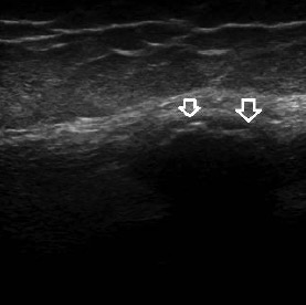
Calcific tendinopathy is most commonly seen around the shoulder joint. Only a few cases of quadriceps calcific tendinopathy (QCT) were reported. This study compares pain, function, clinical examination results, and ultrasonographic findings among primary knee osteoarthritis (KOA) patients with or without ultrasonography-detected QCT. A cross-sectional study was conducted on 214 patients with knee OA. Ultrasonography (US) of knee joints was performed according to the EULAR guidelines. Kellgren-Lawrence radiographic grading was used to score OA. Pain and functional status were assessed using the visual analog scale (VAS), the Health Assessment Questionnaire-II (HAQ-II), and the Western Ontario and McMaster Universities Osteoarthritis Index (WOMAC). QCT was detected in 50 out of 428 knees (11.6%), i.e. in 46 out of 214 patients (21.49%). Most cases of QCT were detected in the following sites: 36 in the vastus lateralis (72%), 10 in the vastus intermedius (20%), and only 4 in the vastus medialis (8%). QCT was found mainly in advanced KOA stages: 44 cases of QCT were found in patients with grade 4 KOA and 6 cases in grade 3 KOA. The presence of QCT showed a statistically significant association (p<0.05) with VAS, HAQ-II, WOMAC subscales, synovitis, and effusion detected by US. In knees with ultrasound-detected QCT, ultrasonographic features of CPPD were found in 31 knees (62%). QCT was found in cases with advanced KOA and mainly with ultrasonographic findings of CPPD disease. QCT could be considered an independent poor prognostic finding regarding pain, functional activity, and response to NSAIDs.

Dimensions

Altmetric

PlumX Metrics

Downloads

Download data is not yet available.

Citations

Speed CA, Hazleman BL. Calcific tendinitis of the shoulder. N Engl J Med. 1999; 340: 1582-1584. DOI: https://doi.org/10.1056/NEJM199905203402011
Bosworth BW. Calcium deposits in the shoulder and subacromial bursitis. A survey of 12, 222 shoulders. JAMA. 1941a; 116: 2477-2482. DOI: https://doi.org/10.1001/jama.1941.02820220019004
Bosworth BW. Examination of the shoulder for calcium deposits. J Bone Joint Surg Am. 1941b; 23: 567-577.
Holt, P.D., Keats, T.E.Calcific tendinitis: a review of the usual and unusual. Skeletal Radiol. 1993; 22: 1-9. DOI: https://doi.org/10.1007/BF00191518
Pereira BP, Chang EY, Resnick DL, Pathria MN. Intramuscular migration of calcium hydroxyapatite crystal deposits involving the rotator cuff tendons of the shoulder: report of 11 patients. Skeletal Radiol. 2016; 45: 97-103. DOI: https://doi.org/10.1007/s00256-015-2255-9
Trujeque L. Patellar whiskers and acute calcific quadriceps tendonitis in a general hospital population. Arthritis Rheum. 1977; 20: 1409-1412. DOI: https://doi.org/10.1002/art.1780200716
Macurak RB, Goldman J A, Hirsh E, et al. Acute calcific quadriceps tendonitis. South Med J. 1980; 73: 322-325. DOI: https://doi.org/10.1097/00007611-198003000-00014
Bhole R, Flynn JC, Marbury TC. Quadriceps tendon ruptures in uremia. Clin Orthop Relat Res.1985; 195: 200-6. DOI: https://doi.org/10.1097/00003086-198505000-00023
Freire V, Moser TP, Lepage-Saucier M. Radiological identification and analysis of soft tissue musculoskeletal calcifications. Insights Imaging 2018; 9: 477-492. DOI: https://doi.org/10.1007/s13244-018-0619-0
Altman R, Asch E, Bloch D, et al. Development of criteria for the classification and reporting of osteoarthritis. Classification of osteoarthritis of the knee. Diagnostic and Therapeutic Criteria Committee of the American Rheumatism Association. Arthritis Rheum. 1986; 29: 1039-1049. DOI: https://doi.org/10.1002/art.1780290816
Bellamy N, Buchanan WW, Goldsmith CH, et al. Validation study of WOMAC: a health status instrument for measuring clinically important patient relevant outcomes to antirheumatic drug therapy in patients with osteoarthritis of the hip or knee. J Rheumatol. 1988; 1833-40.
Wolfe F, Michaud K, Pincus T. Development and validation of the health assessment questionnaire II: a revised version of the health assessment questionnaire. Arthritis Rheum. 2004; 50: 3296-305. DOI: https://doi.org/10.1002/art.20549
Kellgren JH, Lawrence JS. Radiological assessment of osteo-arthrosis. Ann Rheum Dis. 1957;16:494-502. DOI: https://doi.org/10.1136/ard.16.4.494
Möller I, Janta I, Backhaus M, et al. The 2017 EULAR standardised procedures for ultrasound imaging in rheumatology. Ann Rheum Dis. 2017; 76: 1974‐1979. DOI: https://doi.org/10.1136/annrheumdis-2017-211585
Filippou G, Scirè CA, Damjanov N, et al. Definition and reliability assessment of elementary ultrasonographic findings in calcium pyrophosphate deposition disease: a study by the OMERACT calcium pyrophosphate deposition disease ultrasound subtask force. J Rheumatol. 2017;44:1744-1749. DOI: https://doi.org/10.3899/jrheum.161057
Yang BY, Sartoris DJ, Resnick D, Clopton P. Calcium pyrophosphate dihydrate crystal deposition disease: frequency of tendon calcification about the knee. J Rheumatol. 1996; 23: 883-8.
Mohammed R, Kotb H, Amir M, Di Matteo A. Subclinical crystal arthropathy: a silent contributor to inflammation and functional disability in knees with osteoarthritis - an ultrasound study. J Med Ultrasonics. 2019; 46: 137-146. DOI: https://doi.org/10.1007/s10396-018-0912-z

How to Cite

Mortada, M., Kotb, L., & Amer, Y. (2021). Impact of ultrasonography detected quadriceps calcific tendinopathy on pain and function in patients with primary knee osteoarthritis. Reumatismo, 73(2), 111–116. https://doi.org/10.4081/reumatismo.2021.1381