
The place of 18F FDG PET/CT in the management of patients with eosinophilic fasciitis: a case report
All claims expressed in this article are solely those of the authors and do not necessarily represent those of their affiliated organizations, or those of the publisher, the editors and the reviewers. Any product that may be evaluated in this article or claim that may be made by its manufacturer is not guaranteed or endorsed by the publisher.
Accepted: 4 November 2020
Authors
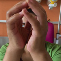
Eosinophilic fasciitis is a rare connective tissue disease with a clinical presentation of scleroderma-like disease. We report a case of a 36-year-old female patient with a 6-month history of progressive stiffness involving her forearms and legs with joint pain. Laboratory examinations showed hypereosinophilia and elevated C-reactive protein. 18F FDG PET/CT showed diffuse and symmetrical increased uptake in the fasciae of the upper and lower limbs, sparing both muscles and fat tissues. Guided biopsy and histologic examination confirmed the diagnosis of eosinophilic fasciitis. 18F FDG PET/CT is of great help in the diagnosis of eosinophilic fasciitis, as it can guide the biopsy where FDG uptake is strongest and also help rule out possible associated neoplasms.
Downloads
Citations


